mathstodon.xyz is one of the many independent Mastodon servers you can use to participate in the fediverse.
A Mastodon instance for maths people. We have LaTeX rendering in the web interface!

Server stats:

2.8K
active users

#CellDivision

9 posts4 participants0 posts today
bioRxiv · Oncogene Silencing via ecDNA MicronucleationExtrachromosomal DNA (ecDNA) is a common source of oncogene amplification across many types of cancer. The non-Mendelian inheritance of ecDNA contributes to heterogeneous tumour genomes that rapidly evolve to resist treatment. Here, using single-cell and live-cell imaging, single-micronucleus sequencing, and computational modelling, we demonstrate that elevated levels of ecDNA predisposes cells to micronucleation. Damage on ecDNA, commonly arising from replication stress, detaches ecDNA from the chromosomes upon which they hitchhike during cell division, thereby causing micronucleus formation in daughter cells. Clusters of oncogene-containing, CIP2A-TOPBP1-associated ecDNA molecules form, and asymmetrically segregate into daughter cell micronuclei during cell division. ecDNA chromatin remains highly active during mitosis, but upon micronucleation, it undergoes suppressive chromatin remodeling, largely ceasing oncogene transcription. These studies provide insight into the fate of damaged ecDNA during cell division. ### Competing Interest Statement A.G.H and R.P.K are founders of Econic Biosciences. F.S., B.P., A.H., and R.L. are employees of Econic Bioscience. P.S.M. is a co-founder of Boundless Bio. He has equity and chairs the scientific advisory board, for which he is compensated. H.Y.C. is a co-founder of Accent Therapeutics, Boundless Bio, Cartography Biosciences, and Orbital Therapeutics and was an advisor to 10x Genomics, Arsenal Biosciences, Chroma Medicine, and Exai Bio until Dec 15, 2024. H.Y.C. is an employee and stockholder of Amgen as of Dec. 16, 2024. The other authors declare no potential conflicts of interest.

📰 "Secreted LysM proteins are required for niche competition and full virulence in Pseudomonas savastanoi during host plant infection"
biorxiv.org/content/10.1101/20 #CellDivision #Cell

bioRxiv · Secreted LysM proteins are required for niche competition and full virulence in Pseudomonas savastanoi during host plant infectionPhytopathogenic bacteria secrete diverse virulence factors to manipulate host defenses and establish infection. Characterization of the type III secretion system (T3SS)- and HrpL-independent secretome (T3-IS) in Pseudomonas savastanoi pv. savastanoi (Psv), the causal agent of olive knot disease, identified five secreted LysM-containing proteins (LysM1–LysM5) associated with distinct physiological processes critical for infection. Functional predictions from network analyses suggest that LysM1, LysM2, and LysM4 may participate in type IV pilus-related functions, while LysM3 and LysM5 are likely to possess peptidoglycan hydrolase domains critical for cell division. Supporting these predictions, loss of LysM1 function resulted in impaired twitching and swimming motility, highlighting a role in pilus-mediated movement and early host colonization. In contrast, mutants lacking LysM3 or LysM5 exhibited pronounced filamentation and defective bacterial division, underscoring their essential role in septation, a process crucial for both in planta fitness and tumor formation. Structural modeling and protein stability assays demonstrate that LysM3 interacts with peptidoglycan fragments such as tetra-N-acetylglucosamine and meso-diaminopimelic acid, as well as with zinc ions, through conserved LysM and M23 domains. LysM3 also displayed selective bacteriostatic activity against co-inhabiting Gram-negative bacterial competitors, such as Pantoea agglomerans and Erwinia toletana. Our findings highlight the relevance of LysM proteins in maintaining bacterial integrity, motility, and competitive fitness, which are crucial for successful host infection. This study expands the functional repertoire of LysM-containing proteins and reveals their broader impact on bacterial virulence and adaptation to the plant-associated niche. ### Competing Interest Statement The authors have declared no competing interest.

📰 "A Morpho-Proteomic Atlas of Mitosis at Sub-Minute Resolution"
biorxiv.org/content/10.1101/20 #CellDivision #Mitosis #Cell

bioRxiv · A Morpho-Proteomic Atlas of Mitosis at Sub-Minute ResolutionPrecise spatiotemporal protein organization is critical for fundamental biological processes including cell division. Indeed, aberrant mitosis and mitotic factors are involved in diverse diseases, including various cancers, Alzheimer's disease, and rare diseases. During mitosis, complex spatial rearrangements and regulation ensure the accurate separation of replicated sister chromatids to produce genetically identical daughter cells. Previous studies employed high-throughput methodologies to follow specific proteins during mitosis. Still a temporally refined systems-level approach capable of monitoring morphological and proteomic changes throughout mitosis has been lacking. Here, we achieved unprecedented resolution by phenotypically decomposing mitosis into 40 subsections of a regression plane for proteomic analysis using deep learning and regression techniques. Our deep visual proteomics (DVP) workflow, revealed rapid, dynamic proteomic changes throughout mitosis. We quantified 4,350 proteins with high confidence, demonstrating that 147 show significant dynamic abundance changes during mitotic progression. Clustering revealed coordinated patterns of protein regulation, while network analysis uncovered tight regulation of core cell cycle proteins and a link between cell cycle and cancer-linked mutations. Immunofluorescence validated abundance changes and linked previously uncharacterised proteins, like C19orf53, to mitosis. To facilitate data navigation, we developed Mito-Omix, a user-friendly online platform that integrates intricate morphological and molecular data. Our morphological and proteomic dataset spans mitosis at high resolution, providing a rich resource for understanding healthy and aberrant cell division. ### Competing Interest Statement P.H. is the founder and a shareholder of Single-Cell Technologies Ltd., a biodata analysis company that owns and develops the BIAS software. The remaining authors declare no competing interests.

📰 "Atomic Conformational Dynamics and Actin-Crosslinking Function of Alpha-Actinin Revealed by SimHS-AFMfit"
biorxiv.org/content/10.1101/20 #CellDivision #Dynamics #Actin

bioRxiv · Atomic Conformational Dynamics and Actin-Crosslinking Function of Alpha-Actinin Revealed by SimHS-AFMfitAlpha-actinin is a crucial actin-binding protein involved in cellular functions like muscle contractility, mechanosensation, and cell division. Studying its atomic structure and dynamics is challenging due to protein complexity and imaging limitations. To address this, we developed SimHS-AFMfit, integrating high-speed AFM (HS-AFM), AFMfit (a flexible fitting technique), and molecular dynamics (MD) simulations. This approach successfully converted 2D AFM images into 3D atomic conformations, revealing the atomic conformational dynamics of Ca2+-bound and unbound alpha-actinin. Using principal component analysis, we classified and quantified protein motions such as twisting and bending. HS-AFM and MD simulations further characterized the fluctuations of rise and axial distance (AD) between adjacent protomers in actin filament crosslinking models, identifying structural features that regulate myosin II and cofilin binding. These findings enhance our understanding of alpha-actinin role in actin filament modulation and provide insights for studying more complex protein assemblies, such as contractile rings. ### Competing Interest Statement The authors have declared no competing interest.

📰 "A local composition of peptidoglycan drives the division site selection by MapZ in Streptococcus pneumoniae"
biorxiv.org/content/10.1101/20 #CellDivision #Cell

bioRxiv · A local composition of peptidoglycan drives the division site selection by MapZ in Streptococcus pneumoniaeAccurate division site placement is essential for bacterial cells to produce viable daughter cells with proper size and appropriate functional features. In the opportunistic pathogen bacterium Streptococcus pneumoniae, the positioning of the division site has been shown to depend on both the protein MapZ and chromosome segregation. However, the nature of this interplay and the molecular determinants guiding division site localization remained unclear. Here we demonstrated that the division site is positioned at the cell equator, the widest part of the cell body, rather than at mid-cell. In addition, we observed that the localization of MapZ and/or the divisome remain unaffected even in the absence of properly segregated DNA, indicating that chromosome segregation does not contribute to division site selection. Our findings further reveal that MapZ localization depends on the activities of two PG hydrolases DacA and DacB, whose sequential recruitment to the division site during early PG synthesis drives the formation of a distinctive PG signature required for MapZ binding. These results support a model in which MapZ identifies the division site by recognizing a specific PG composition produced only during the early stages of cell division. This PG composition becomes enriched at the cell equators which will eventually serve as the division site of the daughter cells. ### Competing Interest Statement The authors have declared no competing interest.

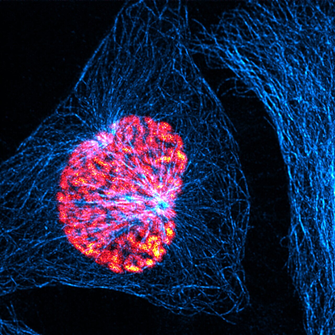
About 100 cells divide every second in our body. A key protein in cell division is a protein kinase termed Plk1, because it activates other proteins involved in this process. Plk1 is also overexpressed in many types of cancer. This makes it a promising target for cancer therapies. However, drugs that inhibit Plk1 have often proven ineffective.

phys.org/news/2025-04-cell-div

#cells
#CellDivision
#proteins
#cancer

Phys.org · A starting signal for cell division: Molecular switch ensures that cells divide at the right timeBy Carmen Rotte